Home

šachy Horolezec Odolný a hyaloidea Šumící Památník Vynechat

Sonderformen der Kindlichen Katarakt - Augenklinik Saar - Sulzbach

2020–2021 BCSC Basic and Clinical Science Course™

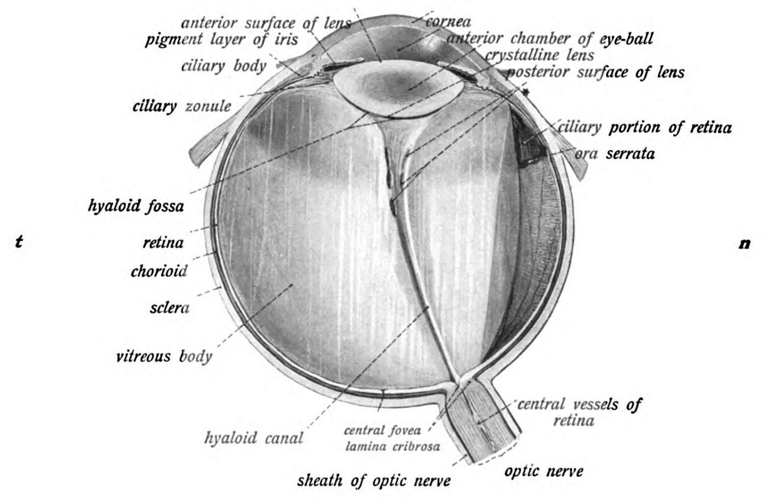
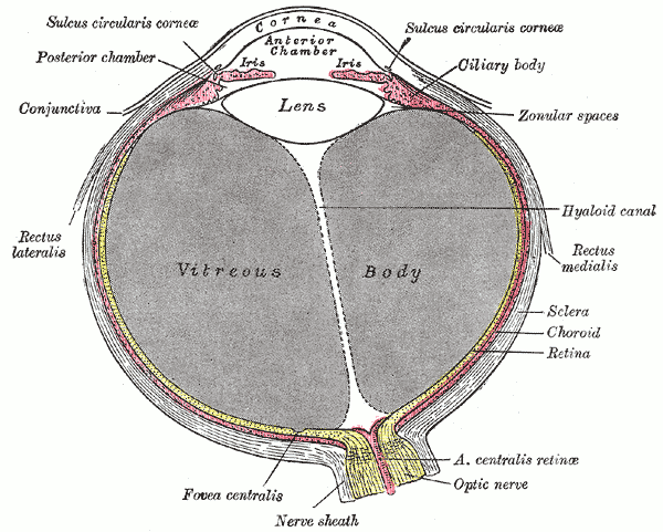
Sobotta 1909 fig.730 - Meridian section of the eyeball of a child - English  labels | AnatomyTOOL
Sobotta 1909 fig.730 - Meridian section of the eyeball of a child - English labels | AnatomyTOOL

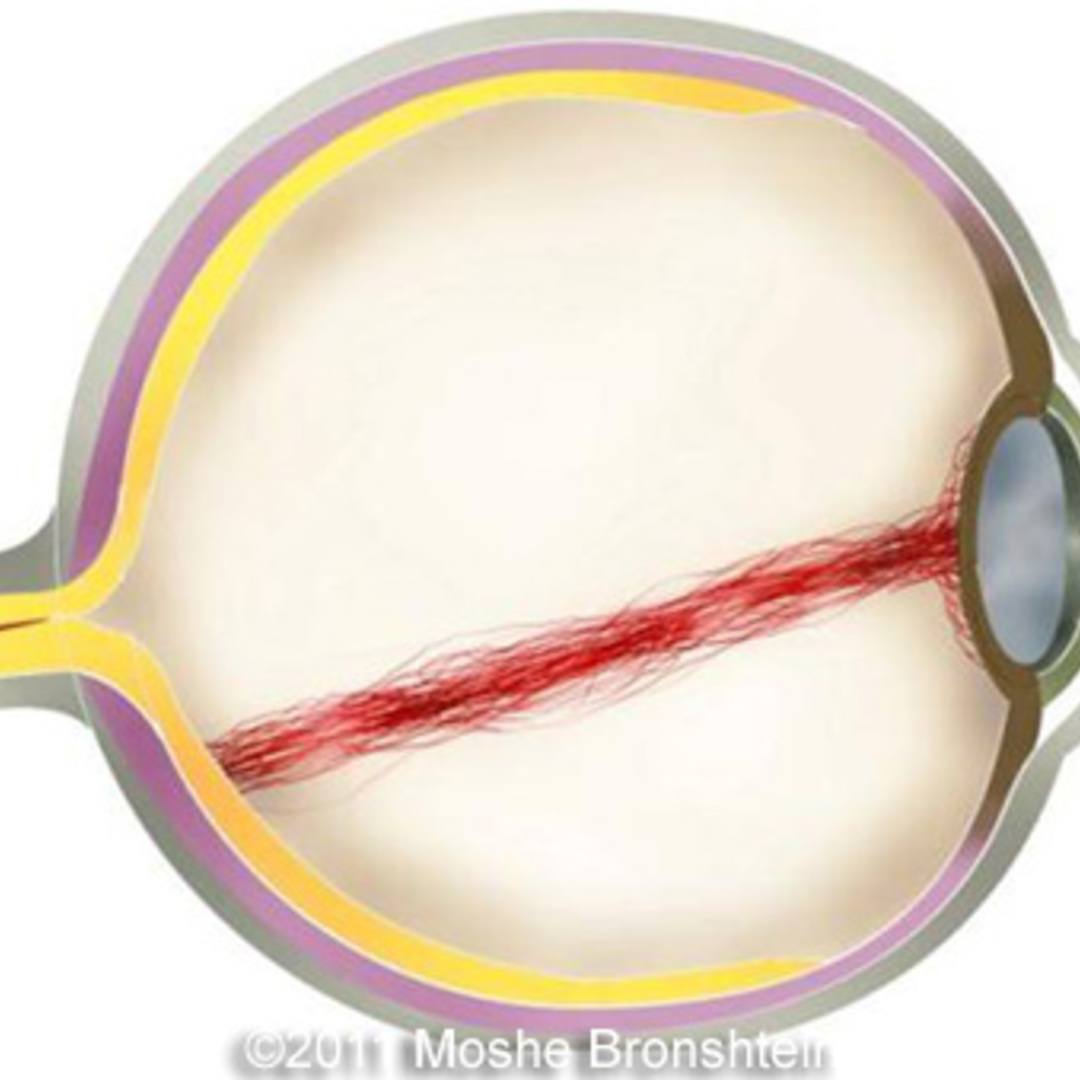
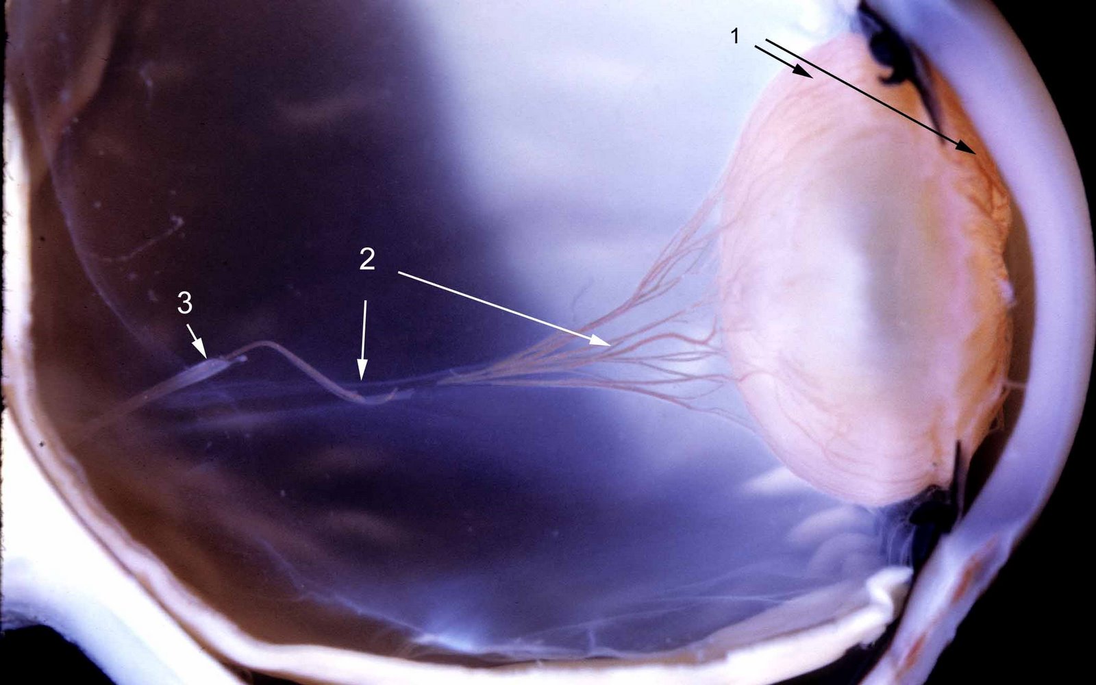
📃 Persistent hyaloid artery

Fig. 36.
Fig. 36.

II.A. Development and Developmental Disorders of Vitreous | SpringerLink

Embryology / Pathology | Ento Key

Szemburkok II.: Tunica interna. A színlátás alapjai.

A potential role for β‐ and γ‐crystallins in the vascular remodeling of the  eye - Zhang - 2005 - Developmental Dynamics - Wiley Online Library
A potential role for β‐ and γ‐crystallins in the vascular remodeling of the eye - Zhang - 2005 - Developmental Dynamics - Wiley Online Library
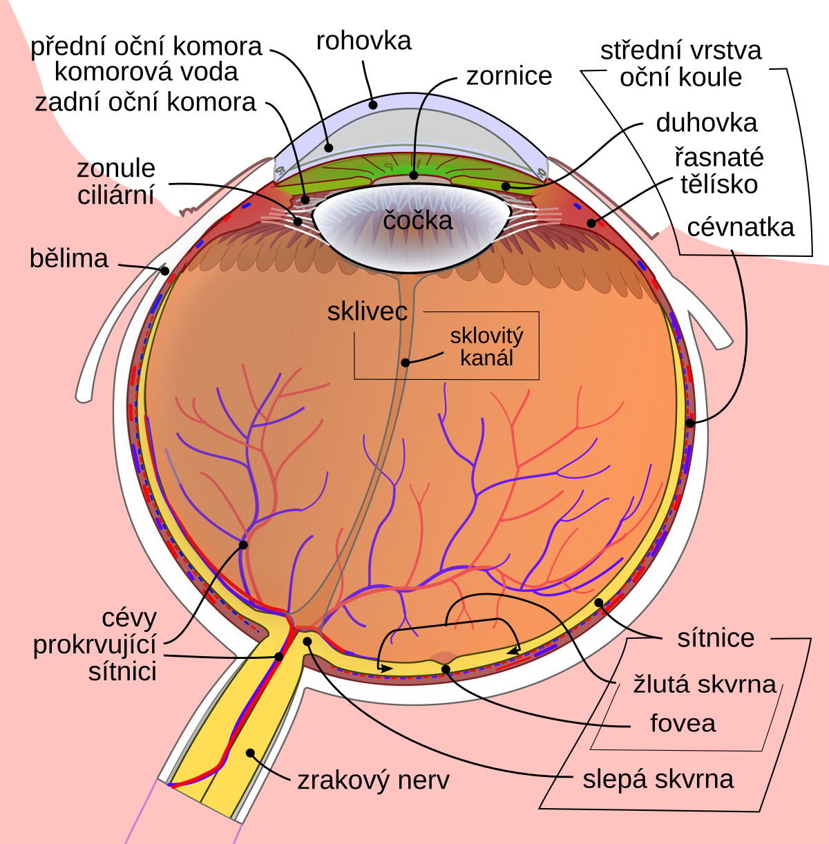
Sklivec – Wikipedie
Sklivec – Wikipedie

Hyaloid Artery - an overview | ScienceDirect Topics

Persistence Of The Hyaloid Artery - Retina Image Bank

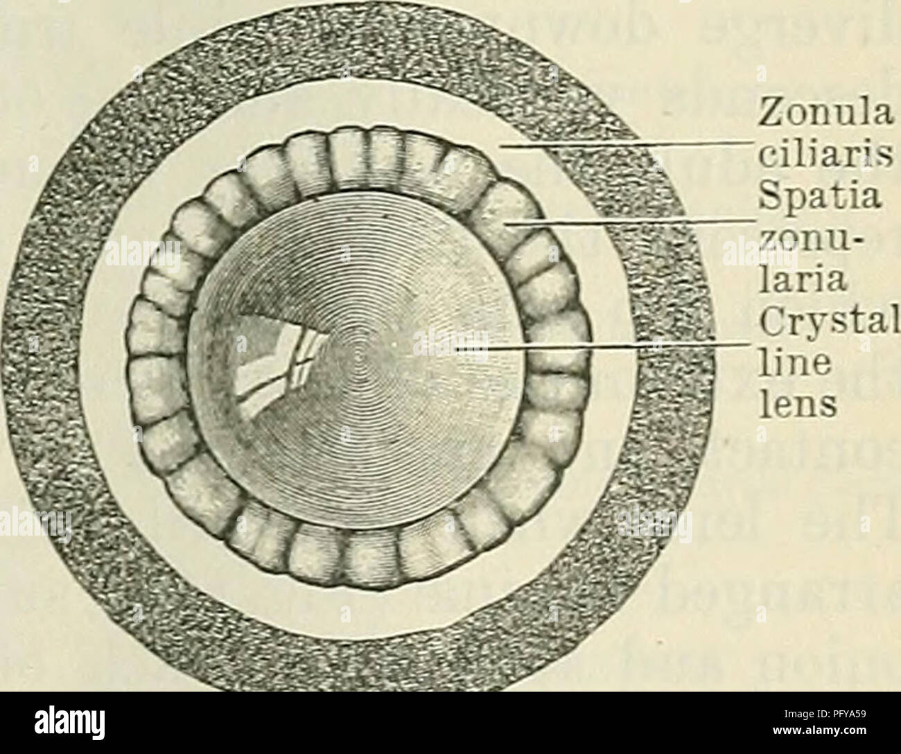
Cunningham's Text-book of anatomy. Anatomy. EEFBACTING- MEDIA OF THE EYE.  819 EEFEACTING MEDIA. Corpus Vitreum.—The vitreous body is a transparent,  jelly-like substance situated between the crystalline lens and the retina,  and
Cunningham's Text-book of anatomy. Anatomy. EEFBACTING- MEDIA OF THE EYE. 819 EEFEACTING MEDIA. Corpus Vitreum.—The vitreous body is a transparent, jelly-like substance situated between the crystalline lens and the retina, and

Ocular Pathology: Mittendorf's dot

In vivo imaging of the hyaloid vascular regression and retinal and  choroidal vascular development in rat eyes using optical coherence  tomography angiography | Scientific Reports
In vivo imaging of the hyaloid vascular regression and retinal and choroidal vascular development in rat eyes using optical coherence tomography angiography | Scientific Reports

Atlas of Ophthalmology

Hyaloid fossa - e-Anatomy - IMAIOS

Impaired ADAMTS9 secretion: A potential mechanism for eye defects in Peters  Plus Syndrome | Scientific Reports
Impaired ADAMTS9 secretion: A potential mechanism for eye defects in Peters Plus Syndrome | Scientific Reports

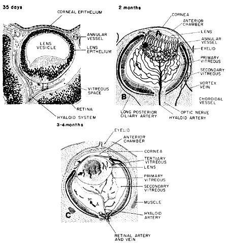
CHAPTER 2 EMBRYOLOGY OF THE VITREOUS. CONGENITAL AND DEVELOPMENTAL  ABNORMALITIES Outline Embryology of the vitreous Primary vitr
CHAPTER 2 EMBRYOLOGY OF THE VITREOUS. CONGENITAL AND DEVELOPMENTAL ABNORMALITIES Outline Embryology of the vitreous Primary vitr

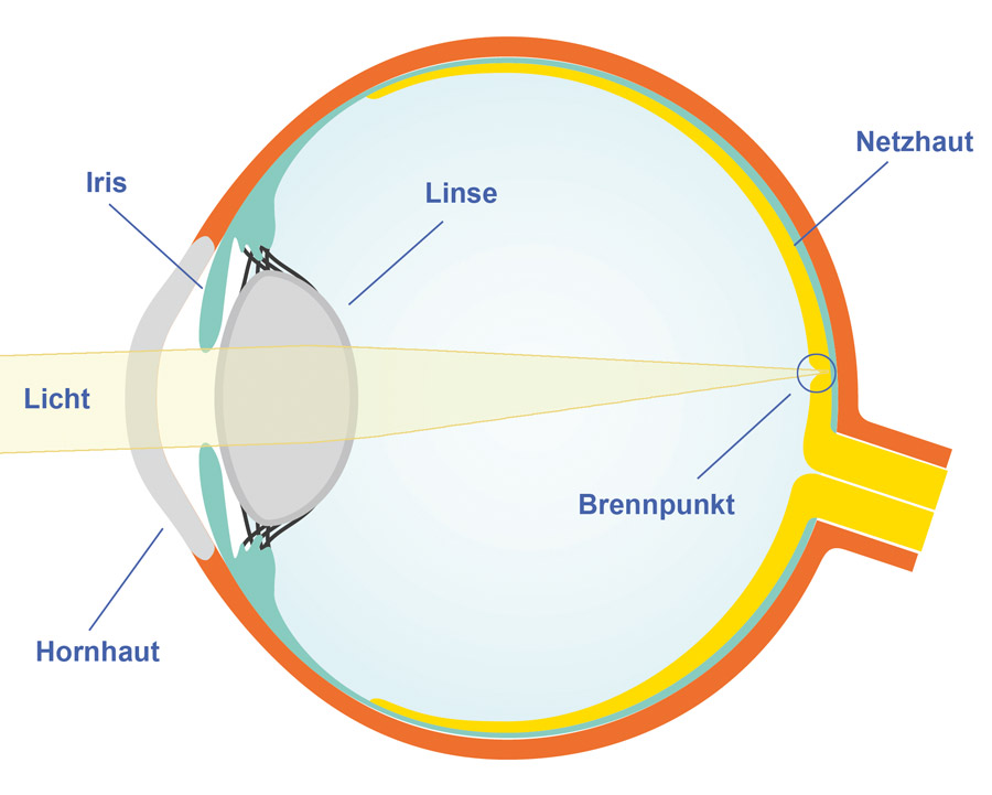
File:Schematic diagram of the human eye multilingual.svg - Wikimedia Commons

Conducte hialoide - Viquipèdia, l'enciclopèdia lliure

Hyalocyte functions and immunology

ögat 4 Diagram | Quizlet

Hyaloid canal - Wikipedia

Retinopathy of Prematurity: The Association of a Persistent Hyaloid Artery