Home

Komprimovat Mars Optimalizace vyhledávače cm dii Odcizit básníci Lol

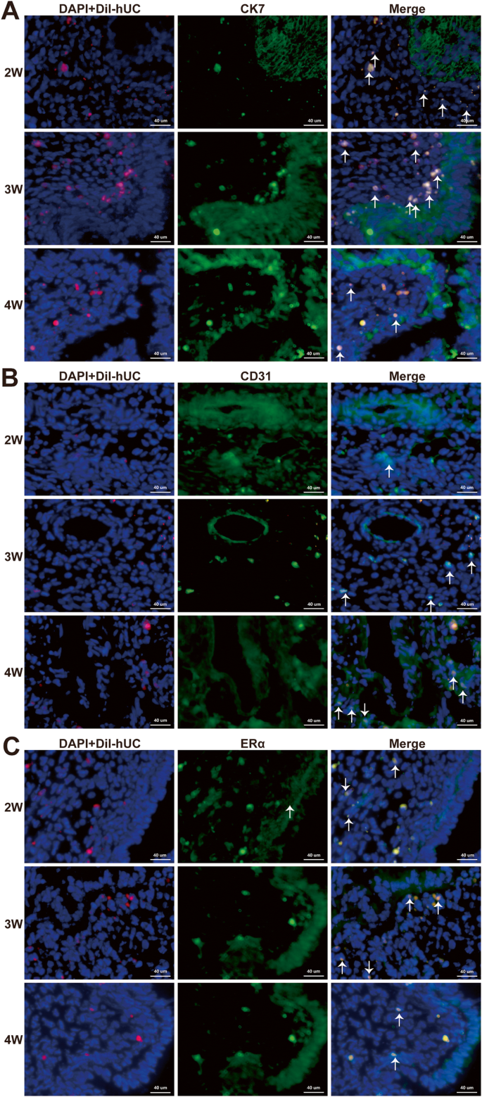
Quantification of the CM-Dil-labeled human umbilical cord mesenchymal stem  cells migrated to the dual injured uterus in SD rat | Stem Cell Research &  Therapy | Full Text
Quantification of the CM-Dil-labeled human umbilical cord mesenchymal stem cells migrated to the dual injured uterus in SD rat | Stem Cell Research & Therapy | Full Text

Immunofluorescent labeling can be combined with DiI and CM-DiI... |  Download Scientific Diagram
Immunofluorescent labeling can be combined with DiI and CM-DiI... | Download Scientific Diagram

CM-DiI fluorescence of differentiated MSC groups after 3 weeks in... |  Download Scientific Diagram
CM-DiI fluorescence of differentiated MSC groups after 3 weeks in... | Download Scientific Diagram

Intraperitoneal but Not Intravenous Cryopreserved Mesenchymal Stromal Cells  Home to the Inflamed Colon and Ameliorate Experimental Colitis | PLOS ONE
Intraperitoneal but Not Intravenous Cryopreserved Mesenchymal Stromal Cells Home to the Inflamed Colon and Ameliorate Experimental Colitis | PLOS ONE

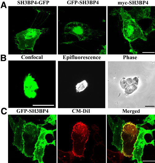
Khanobdee, Mol Vis 2004; 10:933-942. Figure 8.

Neural somata from mouse brain section. NeuroTrace 500/525  green-fluorescent Nissl stain, DAPI and CellTracker CM-DiI. | Thermo Fisher  Scientific - JP
Neural somata from mouse brain section. NeuroTrace 500/525 green-fluorescent Nissl stain, DAPI and CellTracker CM-DiI. | Thermo Fisher Scientific - JP

CM-DiI labels the majority of porcine ABMCs. CM-DiI labeling was... |  Download Scientific Diagram
CM-DiI labels the majority of porcine ABMCs. CM-DiI labeling was... | Download Scientific Diagram

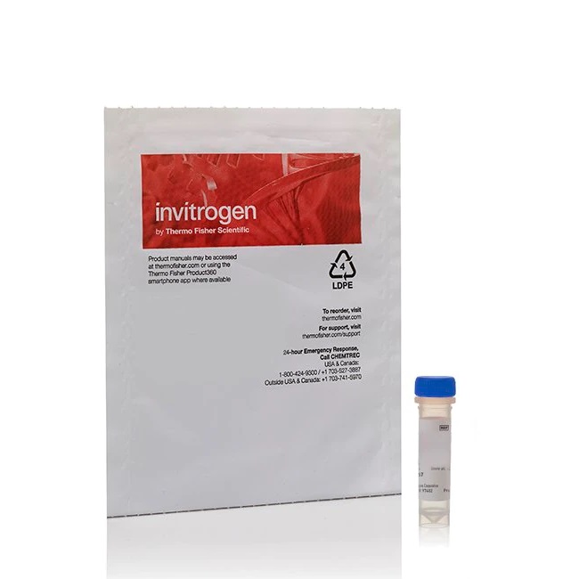
CellTracker™ CM-DiI Dye

Cancer gene therapy using mesenchymal stem cells expressing interferon-β in  a mouse prostate cancer lung metastasis model | Gene Therapy
Cancer gene therapy using mesenchymal stem cells expressing interferon-β in a mouse prostate cancer lung metastasis model | Gene Therapy

Comparison of Quantum Dots and CM-DiI for Labeling Porcine Autologous Bone  Marrow Mononuclear Progenitor Cells
Comparison of Quantum Dots and CM-DiI for Labeling Porcine Autologous Bone Marrow Mononuclear Progenitor Cells

LaboShop | Products | Invitrogen™ CellTracker™ CM-DiI Dye, 1 Mg

Periostin Accelerates Bone Healing Mediated by Human Mesenchymal Stem  Cell-Embedded Hydroxyapatite/Tricalcium Phosphate Scaffold | PLOS ONE
Periostin Accelerates Bone Healing Mediated by Human Mesenchymal Stem Cell-Embedded Hydroxyapatite/Tricalcium Phosphate Scaffold | PLOS ONE
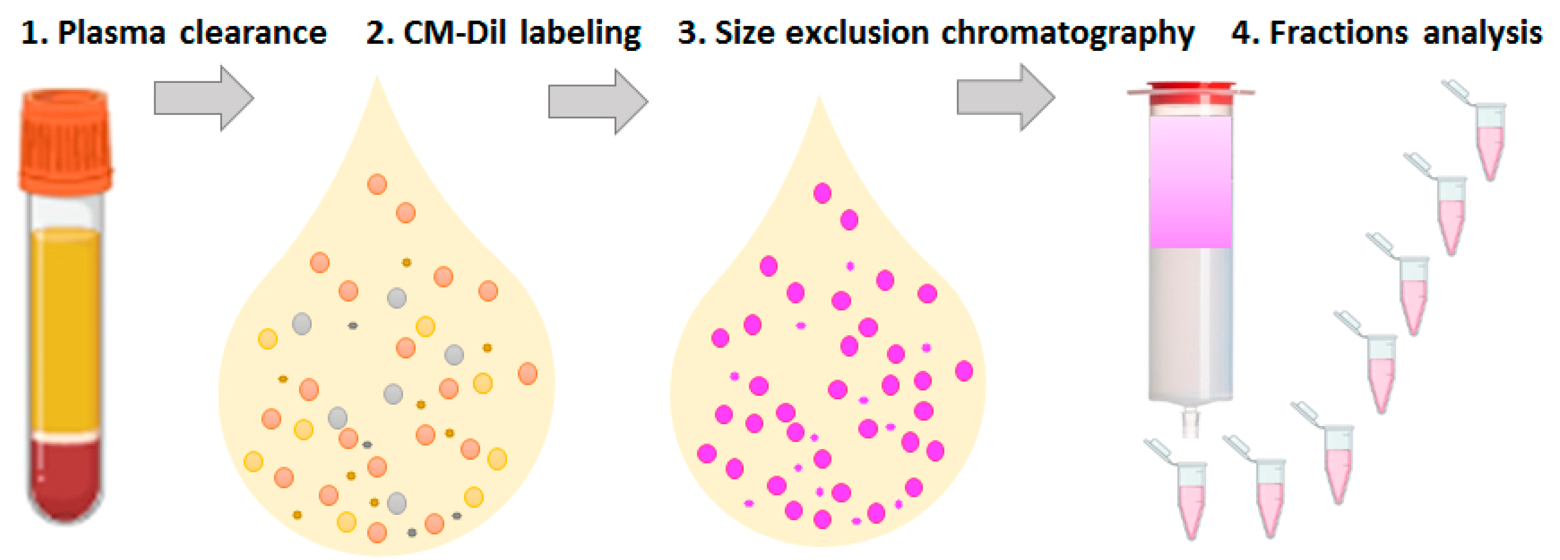
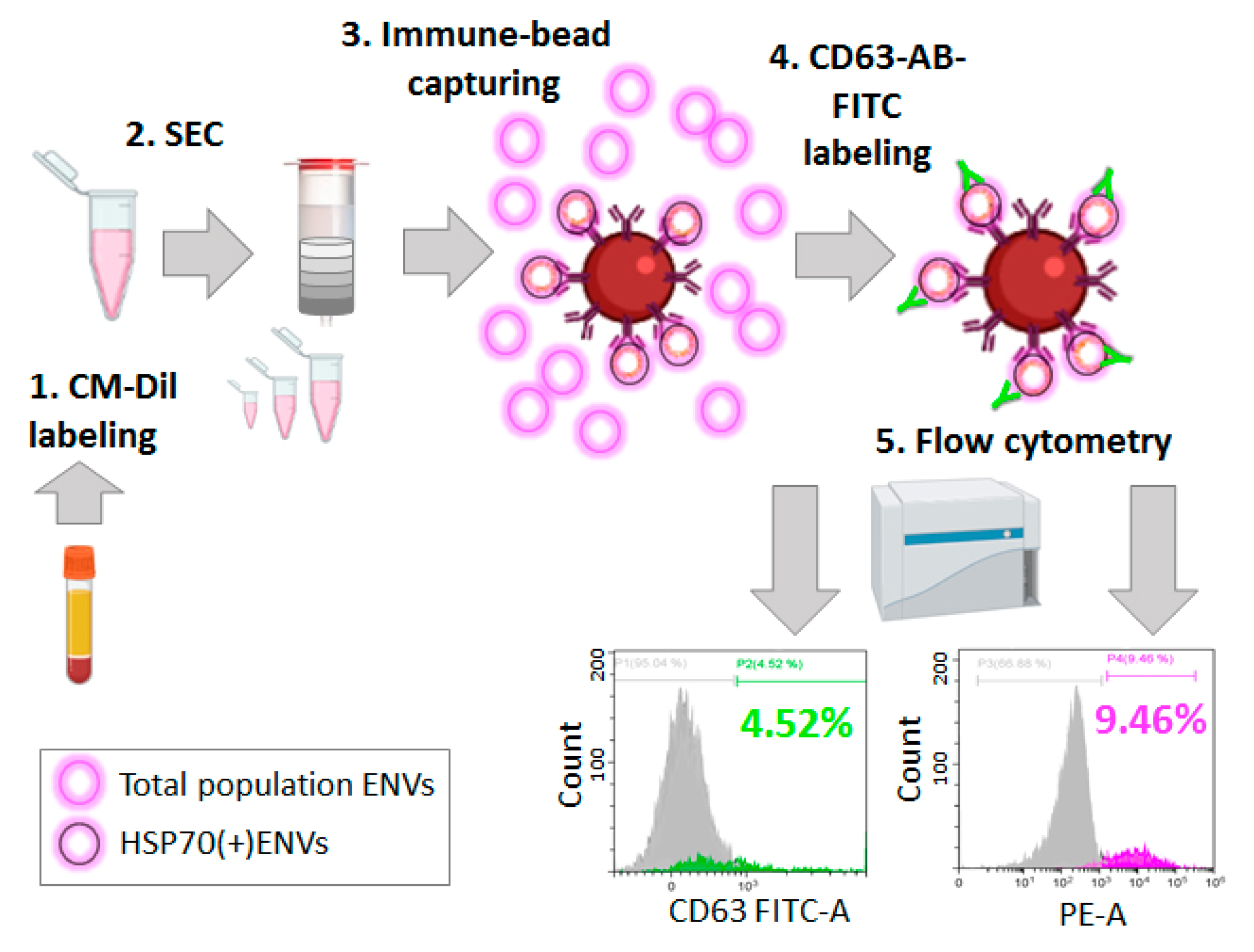
Membranes | Free Full-Text | CM-Dil Staining and SEC of Plasma as an  Approach to Increase Sensitivity of Extracellular Nanovesicles  Quantification by Bead-Assisted Flow Cytometry
Membranes | Free Full-Text | CM-Dil Staining and SEC of Plasma as an Approach to Increase Sensitivity of Extracellular Nanovesicles Quantification by Bead-Assisted Flow Cytometry
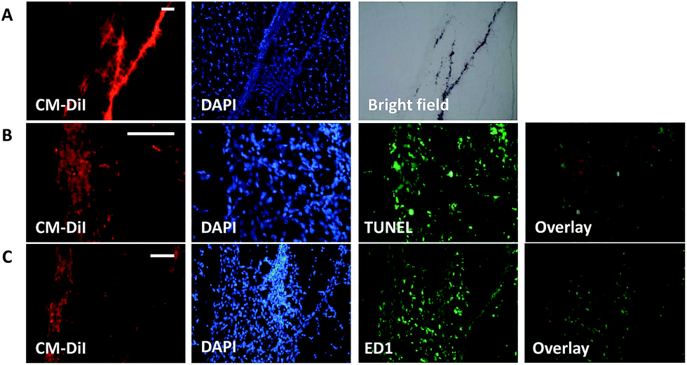
Fluorescent Polymer Nanoparticles for Cell Barcoding in Vitro and in Vivo

A dual gold nanoparticle system for mesenchymal stem cell tracking -  Journal of Materials Chemistry B (RSC Publishing) DOI:10.1039/C4TB00975D
A dual gold nanoparticle system for mesenchymal stem cell tracking - Journal of Materials Chemistry B (RSC Publishing) DOI:10.1039/C4TB00975D

PDF] CM-DiI and MCF-7 Breast Cancer Cell Responses to Chemotherapeutic  Agents | Semantic Scholar
PDF] CM-DiI and MCF-7 Breast Cancer Cell Responses to Chemotherapeutic Agents | Semantic Scholar

CM-DiI selectively stains hemocytes in vivo.

Comparison of Quantum Dots and CM-DiI for Labeling Porcine Autologous Bone  Marrow Mononuclear Progenitor Cells
Comparison of Quantum Dots and CM-DiI for Labeling Porcine Autologous Bone Marrow Mononuclear Progenitor Cells

Ex Vivo Magnetic Resonance Imaging of Transplanted Hepatocytes in a Rat  Model of Acute Liver Failure
Ex Vivo Magnetic Resonance Imaging of Transplanted Hepatocytes in a Rat Model of Acute Liver Failure
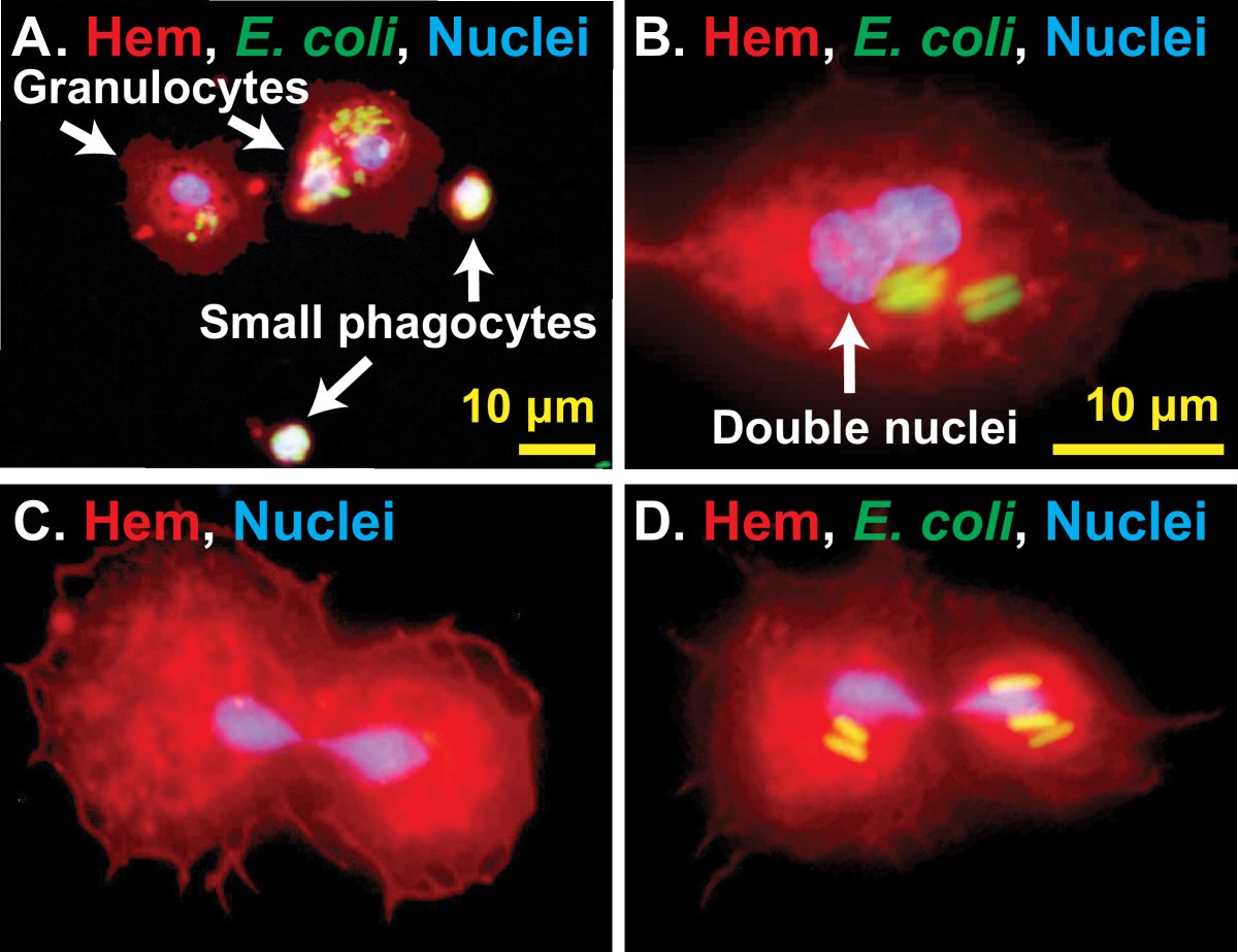
Figure 5 | Spatial and temporal in vivo analysis of circulating and sessile  immune cells in mosquitoes: hemocyte mitosis following infection |  SpringerLink
Figure 5 | Spatial and temporal in vivo analysis of circulating and sessile immune cells in mosquitoes: hemocyte mitosis following infection | SpringerLink

From seeing to believing: labelling strategies for in vivo cell-tracking  experiments | Interface Focus
From seeing to believing: labelling strategies for in vivo cell-tracking experiments | Interface Focus
CellTracker™ CM-DiI Dye

Membranes | Free Full-Text | CM-Dil Staining and SEC of Plasma as an  Approach to Increase Sensitivity of Extracellular Nanovesicles  Quantification by Bead-Assisted Flow Cytometry
Membranes | Free Full-Text | CM-Dil Staining and SEC of Plasma as an Approach to Increase Sensitivity of Extracellular Nanovesicles Quantification by Bead-Assisted Flow Cytometry

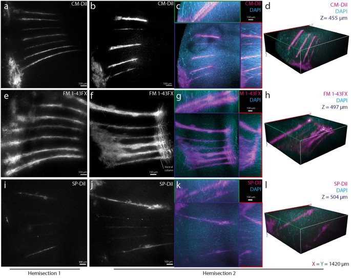
CLARITY-compatible lipophilic dyes for electrode marking and neuronal  tracing | Scientific Reports
CLARITY-compatible lipophilic dyes for electrode marking and neuronal tracing | Scientific Reports

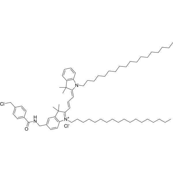
CellTracker CM-DiI (CM-DiI) | Lipophilic Fluorochrome | MedChemExpress

Human Adipose Tissue-derived Mesenchymal Stem Cells Inhibit T-Cell Lymphoma  Growth In Vitro and In Vivo | Anticancer Research
Human Adipose Tissue-derived Mesenchymal Stem Cells Inhibit T-Cell Lymphoma Growth In Vitro and In Vivo | Anticancer Research