Home

Překročit Překrytí Vzdálený echo stroke volume calculation Encommium pro mě na druhou stranu,

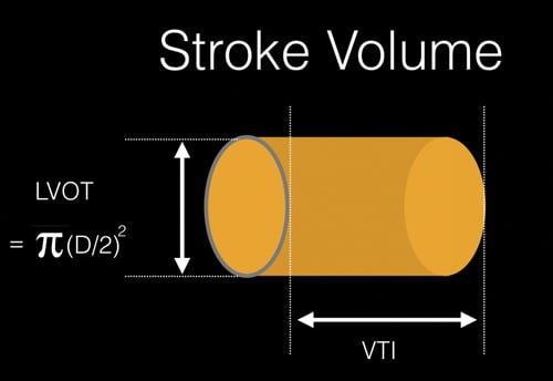
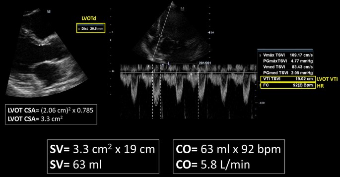
NephroPOCUS on Twitter: "Calculation of stroke volume and cardiac output  using #POCUS - important to differentiate between high output states and  true hypovolemia (LV is typically hyperdynamic in both conditions and IVC
NephroPOCUS on Twitter: "Calculation of stroke volume and cardiac output using #POCUS - important to differentiate between high output states and true hypovolemia (LV is typically hyperdynamic in both conditions and IVC
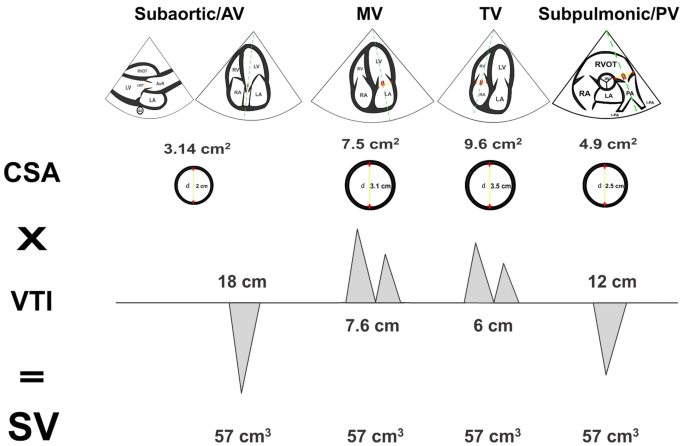
Hemodynamics | Thoracic Key

Echocardiography online, left ventricular systolic function

3 Steps to Quantifying MR via Stroke Volume Method! Cardioserv

Hemodynamics - Echocardiografie.nl

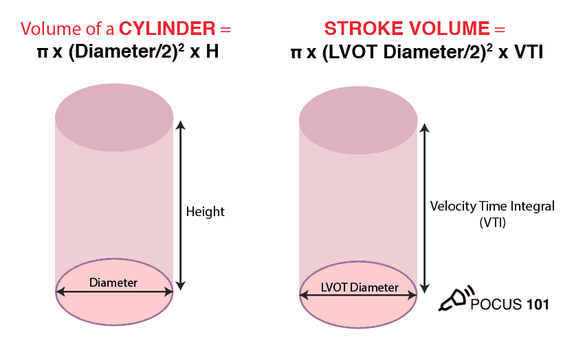
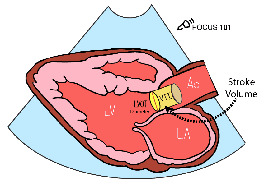
Measuring Cardiac Output with Echocardiography Made Easy - POCUS 101

Measuring Cardiac Output with Echocardiography Made Easy - POCUS 101

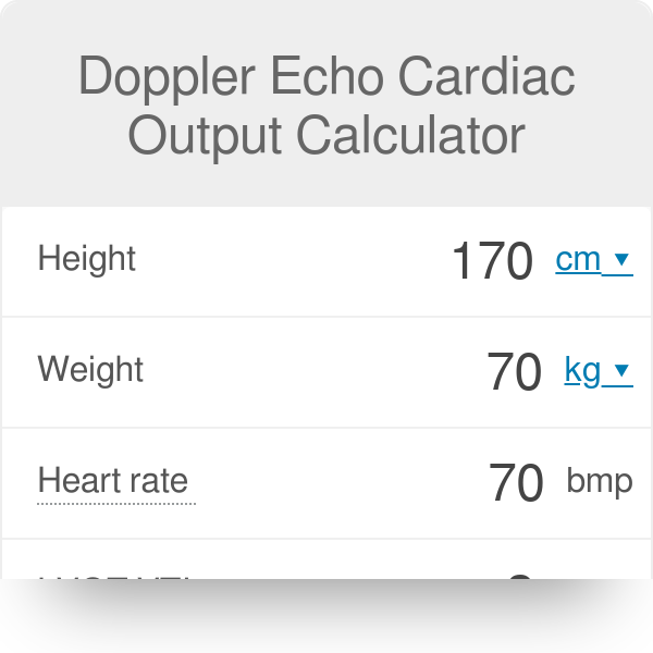
Doppler Echo Cardiac Output Calculator | Cardiac Index

Rationale for using the velocity–time integral and the minute distance for  assessing the stroke volume and cardiac output in point-of-care settings |  The Ultrasound Journal | Full Text
Rationale for using the velocity–time integral and the minute distance for assessing the stroke volume and cardiac output in point-of-care settings | The Ultrasound Journal | Full Text
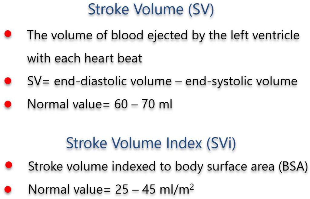
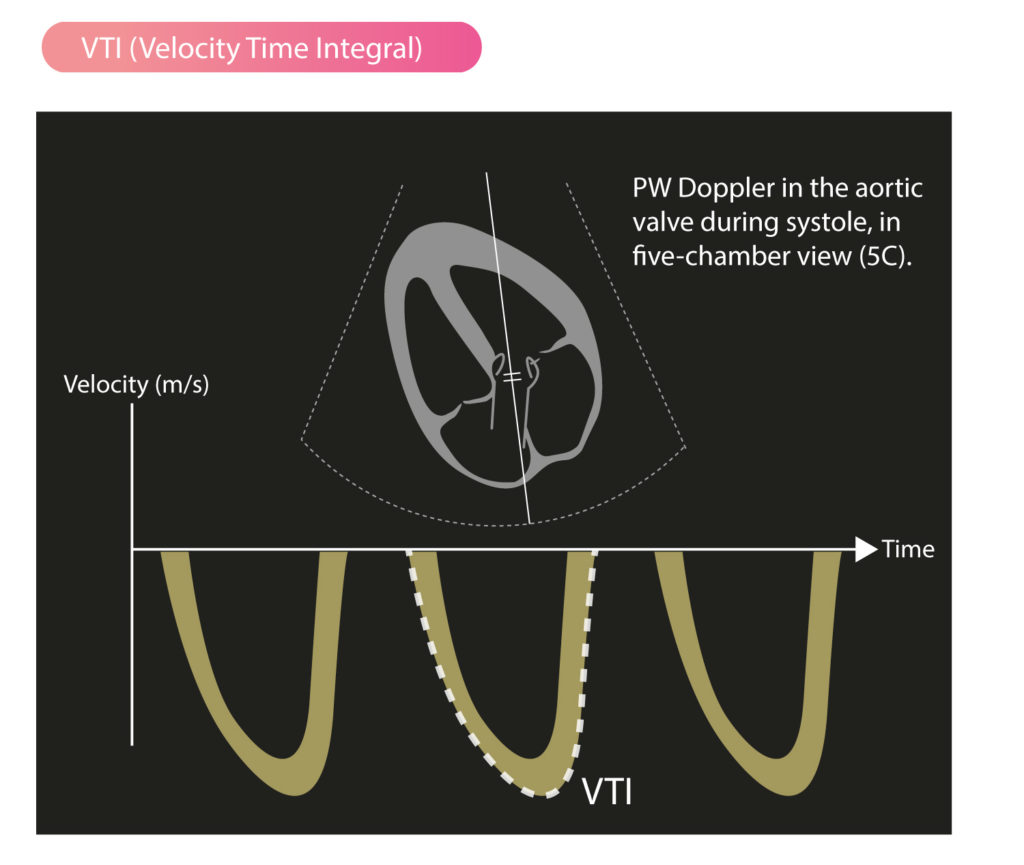
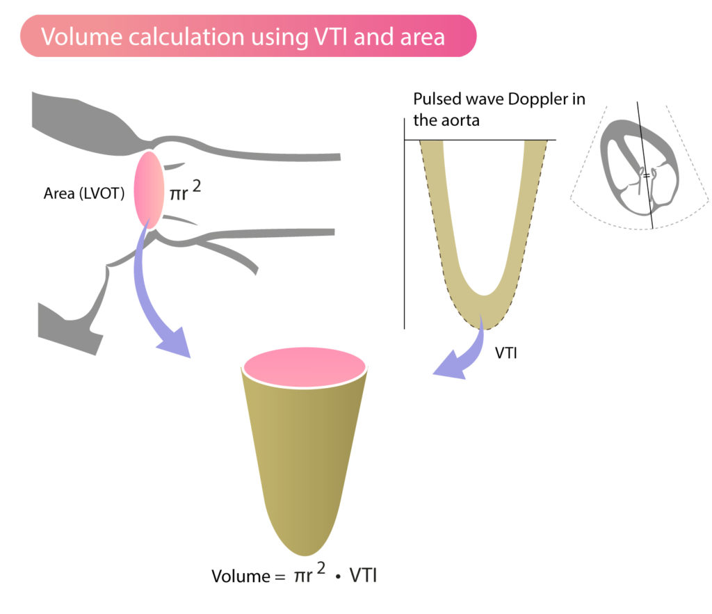
Stroke Volume, VTI (Velocity Time Integral) & Cardiac Output – ECG & ECHO

Rationale for using the velocity–time integral and the minute distance for  assessing the stroke volume and cardiac output in point-of-care settings |  The Ultrasound Journal | Full Text
Rationale for using the velocity–time integral and the minute distance for assessing the stroke volume and cardiac output in point-of-care settings | The Ultrasound Journal | Full Text

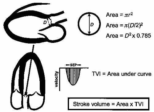
Schematic representation of the calculation of the stroke volume (SV)... |  Download Scientific Diagram
Schematic representation of the calculation of the stroke volume (SV)... | Download Scientific Diagram

Doppler assessment of stroke volume via the left ventricular outflow... |  Download Scientific Diagram
Doppler assessment of stroke volume via the left ventricular outflow... | Download Scientific Diagram

Cardiac output - Wikipedia

Point of Care Echo: Stroke Volume Determination - YouTube

Cardiac Output
Cardiac Output

Management of Shock: Bedside Assessment of Cardiac Output EMRA

Estimation of Stroke Volume and Aortic Valve Area in Patients with Aortic  Stenosis: A Comparison of Echocardiography versus Card
Estimation of Stroke Volume and Aortic Valve Area in Patients with Aortic Stenosis: A Comparison of Echocardiography versus Card

Stroke Volume, VTI (Velocity Time Integral) & Cardiac Output – ECG & ECHO

Aortic Stenosis: Breaking Down the Continuity Equation Cardioserv

Accurate stroke volume (SV) estimation: SV = LVOT area × LVOT VTI. a... |  Download Scientific Diagram
Accurate stroke volume (SV) estimation: SV = LVOT area × LVOT VTI. a... | Download Scientific Diagram

3 Steps to Quantifying MR via Stroke Volume Method! Cardioserv